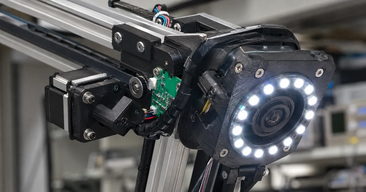
OpenDermは3Dマップを作るオープンソースのロボット
OpenDermは、再現性と解像度がともに高い皮膚の3Dマップを作るオープンソースのロボット撮像システムです。製作者はMarion Lepert(マリオン・レパート)氏で、ハードウェアの設計・ソフトウェア・組み立て文書がすべて公開されています。
位置を合わせたスキャン同士を比較すれば、新しい病変を見つけやすくなり、すでにある病変のわずかな変化にも気づくことができると公式サイトは述べています。同じ場所の前回と今回の画像を並べて確認できるためです。病変とは、皮膚にできた普通とは異なる部分のことです。
用途は研究に限られ、OpenDermは医療機器ではありません。
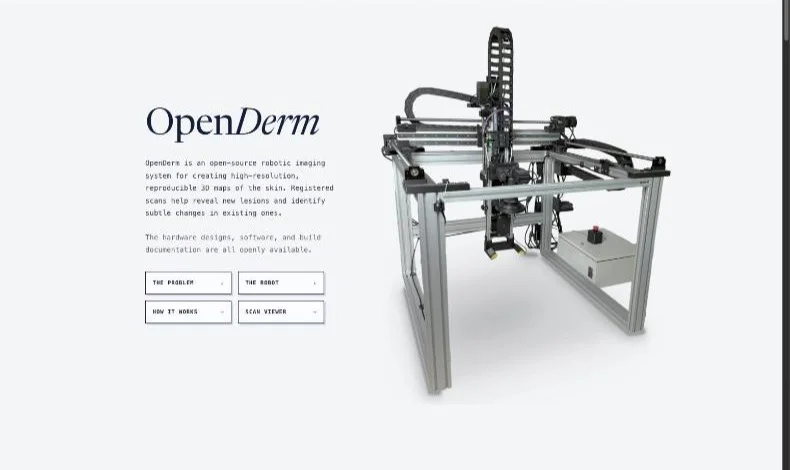
OpenDerm is an open-source robotic imaging system for creating high-resolution, reproducible 3D maps of the skin. Registered scans help reveal new lesions and identify subtle changes in existing ones. / The hardware designs, software, and build documentation are all openly available. / Designed and built by Marion Lepert. / Not a medical device · Research use only — 概要、公開範囲、製作者、および用途の限定に関する記述より公式情報を見る →
78ピクセル/mmの撮影を部品費8,500ドル未満で実現
OpenDermの撮影解像度は1ミリメートルあたり78ピクセルで、ハードウェアの部品費は8,500ドル(約133万2,000円)未満です。 この解像度なら、ほくろの色素網目模様や、そばに生えている毛を1本ずつ見分けられます。
- 撮影解像度:1ミリメートルあたり78ピクセル
- 部品費:ハードウェアで8,500ドル(約133万2,000円)未満
- 費用の比較:市販の全身撮影システムと比較して「ごく一部」
- 公開範囲:完全なオープンソース
市販の全身撮影システムと比較して費用が何分の1になるのか、具体的な数字は公開されていません。解像度についても「入手できるなかで最も高い部類」という表現にとどまります。
OpenDerm captures skin at 78 pixels per millimeter, resolving details such as a mole’s pigment network and nearby individual hairs. / OpenDerm captures skin at 78 pixels per millimeter using less than $8,500 in hardware. It is fully open source and provides some of the highest-resolution imaging available at a fraction of the cost of commercial total-body photography systems. — 撮影解像度、費用、および市販の全身撮影システムとの比較に関する記述より公式情報を見る →
4軸ガントリーとレーザー距離センサーで撮影条件を一定に
OpenDermの本体は主に市販の部品でできており、ガントリー(カメラを取り付けて移動させる門型の骨組み)がカメラを4つの自由度で移動させます。 カメラと照明と距離センサーを取り付けた部分がセンサーヘッドです。3つの直線軸がセンサーヘッドを作業空間内の位置へ移動させ、回転軸のRXがヘッドを傾けてカメラを皮膚の面に合わせます。
OpenDermが持つ4つの自由度(公式サイトによる)
センサーヘッドには次の機材が搭載されています。名称と型番は公式サイトの表記に合わせました。
- カメラ:Canon EOS R7(レンズはRF 100 mmのマクロレンズ)
- 照明:Godox MF-R76のリングフラッシュ(交差偏光フィルター付き)
- 距離の測定:下向きのレーザー距離センサー2つ
交差偏光フィルターは、光の反射によるギラつきを減らします。2つのレーザーセンサーで測った距離をもとに、制御系が皮膚との作業距離とカメラの向きを一定に維持します。
撮影では、撮影地点ごとにロボットがまずレーザーセンサー2つでカメラの角度と作業距離を整えたうえで、高解像度の写真を撮ります。その後の処理で、重なり合う画像を位置合わせして1つの連続した面に再構成するとのこと。
1台のカメラを移動させるのには理由があります。カメラが体に近づいて輪郭に沿って移動すれば、距離・角度・焦点・照明を一定にしたまま、皮膚の表面を高解像度で撮ることができるからです。さらに、安価なアクチュエーター(移動させるための駆動部品)と1台のカメラにとどまるため、固定カメラを大量に並べる構成よりハードウェアの費用を抑えられます。
OpenDerm is built primarily from off-the-shelf parts. / The gantry moves the camera through four degrees of freedom. Three linear axes position the sensor head within the workspace: X travels along the side rails, Y moves across the top beam, and Z sets the camera height. A rotary axis (RX) tilts the sensor head to align the camera with the skin surface. / The sensor head carries a Canon EOS R7 with an RF 100 mm macro lens, a Godox MF-R76 ring flash fitted with a cross-polarization filter that reduces specular glare, and two downward-facing laser distance sensors. The distance measurements allow the control system to maintain a consistent working distance and camera orientation relative to the skin. / At each imaging station, the robot uses two laser sensors to control the camera’s angle and working distance before capturing a high-resolution photograph. The complete pipeline then aligns the overlapping images and reconstructs them as one continuous surface. / By moving close to the body and following its contours, the system can maintain consistent distance, angle, focus, and lighting while capturing high-resolution images across the skin surface. Using a single camera with cheap actuators also reduces hardware cost compared with a large array of fixed cameras. — 市販部品を中心とした構成、4つの自由度、センサーヘッドの中身、撮影の流れ、およびロボット方式を選んだ理由に関する記述より公式情報を見る →
近接ロボット走査は広角の全身撮影より細かいが時間がかかる
公式サイトは広域の皮膚撮影を3つの撮り方に分けて比較しており、OpenDermは近接ロボット走査で撮影します。 速度・詳細さ・費用の組み合わせは、撮り方ごとに異なります。
皮膚の広域撮影における3つの撮り方(公式サイトによる)
| 撮り方 | 速度 | 詳細さ | 費用 | 公式サイトが例に出している製品 |
|---|---|---|---|---|
| 広角の全身撮影 | 秒 | 広角相応 | $$$ | Neko / Canfield VECTRA / DermSpectra / FotoFinder |
| 近接ロボット走査 | 分 | 高解像度 | $$ | SquareMind / iToBoS / OpenDerm |
| スマートフォン誘導撮影 | 手動 | ばらつく | $ | SkinIO / MoleMap / Miiskin |
近接ロボット走査では、高解像度のカメラをロボットアームやガントリーに取り付け、距離と視野角を制御しながら体の表面に沿って移動させます。領域ごとに撮影するので、1台のカメラでも離れた位置からの広角撮影よりずっと細かく写せ、必要な空間とカメラ機材も少なくなります。
ただし撮影には時間がかかります。体全体を一度に撮らず、部分ごとに順番に走査していくためです。
全身撮影はまだ発展途上の分野で、米国では臨床で使える機会が限られています。多くのシステムは実験段階か開発中です。
Total-body photography remains an emerging field, with limited clinical availability in the United States and many systems still experimental or under development. / A high-resolution camera mounted on a robotic arm or gantry moves close to the skin and systematically across the body while maintaining a controlled distance and viewing angle. Imaging the skin region by region allows a single camera to capture much finer detail than a distant wide-field system, while requiring less space and camera hardware. The trade-off is speed: the body must be scanned sequentially rather than all at once. / SecondsWide-field detail$$$ / Examples: Neko · Canfield VECTRA · DermSpectra · FotoFinder / MinutesHigh-resolution detail$$ / Examples: SquareMind · iToBoS · OpenDerm / ManualVariable detailNo dedicated hardware$ / Examples: SkinIO · MoleMap · Miiskin — この分野の現状、近接ロボット走査方式の特徴と代償、および3つの方式のカードに記された速度・詳細さ・費用・例に関する記述より公式情報を見る →
メラノーマは新規病変が70%、既存のほくろ内が約30%
公式サイト冒頭の問題提起には、米国のメラノーマ(皮膚がんの一種)に関する数値が3つ書かれています。 数値は公式サイトの記載をそのまま転記したもので、当サイトによる医学的な判断は加えていません。
- 米国では毎年10万人を超える人がメラノーマと診断される
- 5年相対生存率は早期に見つかればほぼ100%で、体の離れた部位に達した後では約34%
- メラノーマの約30%は既存のほくろ内に生じ、70%は新しい病変として見られる
最初に見られる兆候は、ほくろの縁のミリ単位の変化にとどまる場合もあります。ほくろの縁のわずかな変化に気づくには、皮膚が以前どういう状態だったかを正確に記録しておかなければなりません。全身撮影の改善に必要とされるのは次の3点です。
- 高解像度の撮像(全身撮影システムの多くは広い視野で撮るため、細部が粗くなる)
- 経時的なデータセット
- 頻繁に、手軽に受けられる走査
More than 100,000 Americans are diagnosed with melanoma each year. This form of skin cancer has a five-year relative survival rate of nearly 100% when detected early, but about 34% after it reaches distant parts of the body. / Approximately 30% of melanomas develop within an existing mole, while 70% appear as new lesions. / In either case, the earliest visible sign may be small: a millimeter-scale change at the edge of a mole, a localized shift in its color or structure, or a new spot only a few millimeters across. Detecting changes at this scale requires a precise record of the skin’s prior appearance. / Improving total-body imaging requires progress in three areas: / Higher Resolution Imaging / Many existing total-body photography systems use wide fields of view to capture large areas of skin, but at the expense of fine detail. / Longitudinal datasets / Frequent, accessible scanning — 米国のメラノーマの数値、最初に見える兆候の小ささ、および改善が必要とされる3領域に関する記述より公式情報を見る →
皮膚がん検出AIの学習には同じ皮膚の経時画像が不足している
経時画像とは、同じ皮膚を時間をおいて繰り返し撮った画像のことです。 皮膚がん検出AIの学習データには、この経時画像が不足しています。
皮膚がん検出AIモデルの多くは、既に疑わしいと判断された病変を1回だけ撮った画像で学習しています。したがって、メラノーマが診察で見つかる前にどう生じてどう進むのかが分かる画像は、データセットにほとんど含まれません。より早い兆候をAIに認識させるには、高解像度の経時画像が必要になります。
公式サイトは、全身撮影がもっと手頃で広く使うことができるものになるべきだとしています。リスクの高い患者は同じ条件のスキャンを頻繁に受けられるべきだとも述べています。
Most AI models for skin-cancer detection are trained on isolated images of lesions already identified as suspicious. These datasets therefore provide little evidence of how melanomas first emerge and evolve before drawing clinical attention. Training models to recognize earlier signs of melanoma will require repeated, high-resolution images of the same skin over time. / Total-body photography must become more affordable and widely available. Patients at high risk should be able to receive frequent, standardized scans. — 既存の学習データセットの限界、必要とされるデータの性質、および普及の必要性に関する記述より公式情報を見る →
ライセンスはMITとCERN-OHL-P-2.0の2種類
OpenDermのライセンスは2種類で、ソフトウェアがMIT License、ハードウェアがCERN-OHL-P-2.0です。ハードウェア側の正式名は、CERN Open Hardware Licence Version 2 - Permissiveといいます。設計ファイル・回路図・部品表・組み立て文書が、このライセンスの対象となります。
オープンソースハードウェアとして公開されている範囲は、部品表・CADの組み立て図・電気および安全の回路図・組み立て手順書です。ソースはGitHubのリポジトリにあり、READMEにはシステム構成(System architecture)・インストール方法(Install)・ライセンス(License)などの説明があります。
組み立てに必要な資料はすべて公開されています。ただし、OpenDermは医療機器ではなく、用途は研究に限られます。
The complete bill of materials, CAD assembly, electrical and safety schematics, and step-by-step build instructions are publicly available. / OpenDerm software is available under the MIT License. The hardware design files, schematics, bill of materials, and build documentation are available under the CERN Open Hardware Licence Version 2 - Permissive (CERN-OHL-P-2.0). / Not a medical device · Research use only — 公開されている資料の範囲、ソフトウェアとハードウェアそれぞれのライセンス、および用途の限定に関する記述より公式情報を見る →
FAQ
- OpenDermとは何ですか?
- 皮膚の高解像度な3Dマップを再現性のある方法で作るための、オープンソースのロボット撮像システムです。位置を合わせたスキャン同士を比較すると、新しい病変を見つけやすくなり、既にある病変のわずかな変化にも気づくことができるとのこと。ハードウェアの設計・ソフトウェア・組み立て文書がすべて公開されています。
- 解像度と部品費はどのくらいですか?
- 1ミリメートルあたり78ピクセルで撮影し、部品費は8,500ドル(約133万2,000円)未満とされています。市販の全身撮影システムと比較してごく一部の費用で、入手できるなかでは最も高い部類の解像度が得られると公式サイトは述べています。
- ライセンスは何ですか?
- ソフトウェアはMIT License、ハードウェアの設計ファイル・回路図・部品表・組み立て文書はCERN Open Hardware Licence Version 2 - Permissive(CERN-OHL-P-2.0)で提供されています。部品表・CADの組み立て図・電気および安全の回路図・組み立て手順書の資料も、すべて公開されています。
- なぜAIの話として取り上げられるのですか?
- 公式サイトが課題のひとつとして学習データを指摘しているからです。皮膚がん検出AIモデルの多くは、既に疑わしいと判断された病変を1回だけ撮った画像で学習しています。したがって、メラノーマが診察で見つかる前にどう生じてどう進むのかが分かる画像は、学習データにほとんど含まれません。より早い段階の兆候を認識させるには、同じ皮膚を繰り返し撮った高解像度の画像が必要だというのが公式サイトの指摘です。
関連する記事
 Apple Intelligenceをオフにする設定と、AIモデルの削除方法macOS 27でApple Intelligenceを機能ごとにオフにする設定と、AIモデルを削除して容量を空ける方法を解説します。Appleの説明には削除の手順がないため、モデルまで削除できる無料ツール「RemoveMacAI」の使い方と、導入前に把握すべき点、元に戻す方法を確認できます。
 Cyber Missionとは?重要インフラをAIで守る計画に日立も参加Anthropicが2026年10月8日に発表した「Cyber Mission」は、重要インフラとオープンソースを守る長期の取り組みです。日立など11社と連携する重要インフラ向けのCIDPと、OSSの脆弱性を無料で調べるOSS Scannerが第1弾です。
 NVIDIAのHugging Face買収、米司法省が競争への影響を調査ロイターは2026年10月28日、NVIDIAによる129億ドル(約1兆9,890億円)のHugging Face買収について、米司法省がAI分野の競争への影響を調査していると報じました。買収はNVIDIAが9月3日に発表し、完了は2027年前半の見込みです。
 Langfuseとは?導入方法とDockerでの構築・料金と無料枠の上限LangfuseはAIアプリの実行記録・プロンプト管理・評価を、まとめて使うことができるオープンソースです。Docker Composeなら約5分で起動できます。クラウド版は無料から月2,499ドル(約39万8,300円)までの4プランです。無料のHobbyプランは月50,000ユニットまで使うことができます。