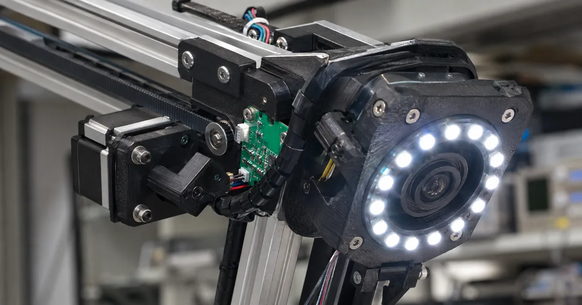
Skin imaging for melanoma checks can use wide-field total-body photography or guided smartphone imaging, and each gives something up. The site says many wide-field systems lose fine detail, and it lists smartphone detail as variable. OpenDerm, an open-source robot designed and built by Marion Lepert, takes a different route: it moves one camera close to the skin and builds a 3D map from many sharp photographs.
The project publishes a capture resolution of 78 pixels per millimeter, a hardware cost under $8,500, and open licenses for both software and hardware. It is a research tool and not a medical device.
What OpenDerm is: an open-source robot for skin imaging
OpenDerm is a robotic imaging system that photographs skin at close range and combines the photos into a high-resolution 3D map of the skin. Marion Lepert designed and built it, and the hardware designs, software, and build documentation are openly available.
OpenDerm is imaging hardware, not a skin analysis app. It takes pictures and builds maps, and the project site describes no model that interprets the images.
The map is meant to be repeatable. Images taken at different times and aligned to each other (the site calls these registered scans) help reveal new lesions and subtle changes in existing ones. The site also marks the project as not a medical device and for research use only.
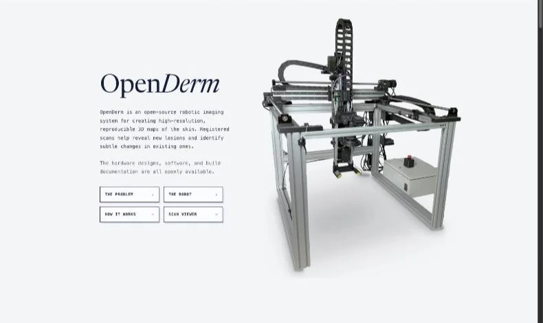
OpenDerm is an open-source robotic imaging system for creating high-resolution, reproducible 3D maps of the skin. Registered scans help reveal new lesions and identify subtle changes in existing ones. / The hardware designs, software, and build documentation are all openly available. / Designed and built by Marion Lepert. / Not a medical device · Research use only — From the statements on what the project is and publishes, on who built it, and on the limits of its intended useView official source →
Why early melanoma detection depends on better imaging
The melanoma figures the project cites
The project starts from melanoma, a form of skin cancer, and reports that more than 100,000 Americans are diagnosed each year. Five-year relative survival is nearly 100% when the cancer is found early, but about 34% once it has reached distant parts of the body. The site's first footnote cites the National Cancer Institute's SEER Cancer Stat Facts: Melanoma of the Skin. The site adds that about 30% of melanomas develop within an existing mole while 70% appear as new lesions.
More than 100,000 Americans are diagnosed with melanoma each year. This form of skin cancer has a five-year relative survival rate of nearly 100% when detected early, but about 34% after it reaches distant parts of the body. / Approximately 30% of melanomas develop within an existing mole, while 70% appear as new lesions. / National Cancer Institute. SEER Cancer Stat Facts: Melanoma of the Skin. — From the incidence, survival, and new-lesion figures and the first footnoteView official source →
Three areas that need progress
Total-body photography (photographing the whole skin surface) can provide a record of the skin over time, but access stays very limited. The site gives two reasons: high costs, and limited clinical evidence that existing systems reliably catch melanoma earlier than dermatologists. The project names three areas where total-body imaging needs to improve:
- Higher-resolution imaging. Many existing total-body photography systems use wide fields of view to cover large areas of skin, at the expense of fine detail.
- Longitudinal datasets (images of the same skin repeated over time). Most AI models for skin cancer detection are trained on isolated images of lesions already flagged as suspicious, and those datasets show little of how melanomas first emerge and evolve.
- Frequent, accessible scanning. Total-body photography has to become more affordable and widely available, and patients at high risk should be able to get frequent, standardized scans.
The project says a robotic imaging system offers a practical way to meet these requirements. A camera that moves in close and follows the body's contours can keep distance, angle, focus, and lighting consistent, and one camera on cheap actuators costs less than a large array of fixed cameras. The project's design is public, and its code is on GitHub.
Total-body photography can provide this longitudinal record, but access to such systems remains very limited due to high costs and limited clinical evidence that existing systems reliably catch melanoma earlier than dermatologists / Improving total-body imaging requires progress in three areas: / Many existing total-body photography systems use wide fields of view to capture large areas of skin, but at the expense of fine detail. / A robotic imaging system offers a practical way to meet these requirements. — From the passages on total-body photography access, on wide-field detail, and on the robotic approachView official source →
How robotic scanning compares with total-body photography
Three approaches compared
The site sorts imaging of large skin areas into three approaches, each trading speed, detail, consistency, footprint, and cost differently. The three-approaches table gives the speed, detail, and cost labels it assigns and the examples it lists.
Three approaches to imaging large areas of skin (per the official site)
| Approach | Speed | Detail | Cost | Examples given |
|---|---|---|---|---|
| Wide-field total-body photography | Seconds | Wide-field detail | $$$ | Neko / Canfield VECTRA / DermSpectra / FotoFinder |
| Close-range robotic scanners | Minutes | High-resolution detail | $$ | SquareMind / iToBoS / OpenDerm |
| Guided smartphone imaging | Manual | Variable detail | $ | SkinIO / MoleMap / Miiskin |
Wide-field systems photograph the body from a distance, and the site says many existing ones give up fine detail to cover large areas of skin. Guided smartphone imaging needs no dedicated hardware, but the site lists its speed as manual and its detail as variable.
Three main approaches have emerged for imaging large areas of skin: wide-field total-body photography, close-range robotic scanning, and guided smartphone imaging. Each involves different trade-offs in speed, detail, consistency, footprint, and cost. / Examples: Neko · Canfield VECTRA · DermSpectra · FotoFinder / Examples: SquareMind · iToBoS · OpenDerm / Examples: SkinIO · MoleMap · Miiskin — From the overview of the approaches and the examples listed in the Approach 01 to 03 cardsView official source →
Close-range scanning trades speed for detail
Close-range robotic scanning puts a high-resolution camera on a robotic arm or a gantry (a frame that carries the camera along rails). The camera moves near the skin and works across the body region by region, holding its distance and viewing angle under control. One camera can then record much finer detail than a wide-field system standing back, and it needs less space and less camera hardware.
The cost is speed. The body is scanned in sequence instead of all at once, and the site lists close-range robotic scanners at minutes where wide-field systems take seconds. Total-body photography as a whole is also still an emerging field: clinical availability in the United States is limited, and many systems are experimental or in development.
Total-body photography remains an emerging field, with limited clinical availability in the United States and many systems still experimental or under development. / A high-resolution camera mounted on a robotic arm or gantry moves close to the skin and systematically across the body while maintaining a controlled distance and viewing angle. Imaging the skin region by region allows a single camera to capture much finer detail than a distant wide-field system, while requiring less space and camera hardware. The trade-off is speed: the body must be scanned sequentially rather than all at once. — From the statements on the state of the field and on the characteristics and trade-off of close-range robotic scanningView official source →
How the robot builds a 3D skin map
OpenDerm turns 3D skin imaging into a sequence of steps: move to a station, set the camera, take a photograph, then merge all the photographs into one surface. The moving frame, the sensor head and the single camera each handle one part of that sequence.
Four axes built from off-the-shelf parts
OpenDerm is built mostly from off-the-shelf parts, meaning ready-made components rather than custom ones. The camera rides on a gantry that moves with four degrees of freedom. Three linear axes place the sensor head in the workspace (X along the side rails, Y across the top beam, Z for camera height), and a rotary axis called RX tilts the head so the camera faces the skin surface.
OpenDerm's four degrees of freedom (per the official site)
OpenDerm is built primarily from off-the-shelf parts. / The gantry moves the camera through four degrees of freedom. Three linear axes position the sensor head within the workspace: X travels along the side rails, Y moves across the top beam, and Z sets the camera height. A rotary axis (RX) tilts the sensor head to align the camera with the skin surface. — From the statements on the off-the-shelf construction and on the four degrees of freedomView official source →
What the sensor head carries
The sensor head carries a Canon EOS R7 camera with an RF 100 mm macro lens, a Godox MF-R76 ring flash with a cross-polarization filter, and two downward-facing laser distance sensors. The filter reduces specular glare, the shiny reflection off the skin surface. The laser readings let the control system hold the working distance and camera orientation steady relative to the skin.
At each imaging station the robot uses the two laser sensors to set the camera angle and working distance, then takes a high-resolution photograph. Afterward, the pipeline aligns the overlapping photographs and rebuilds them as one continuous surface.
The sensor head carries a Canon EOS R7 with an RF 100 mm macro lens, a Godox MF-R76 ring flash fitted with a cross-polarization filter that reduces specular glare, and two downward-facing laser distance sensors. The distance measurements allow the control system to maintain a consistent working distance and camera orientation relative to the skin. / At each imaging station, the robot uses two laser sensors to control the camera’s angle and working distance before capturing a high-resolution photograph. The complete pipeline then aligns the overlapping images and reconstructs them as one continuous surface. — From the statements on what the sensor head carries and on the capture flowView official source →
Why the design uses one moving camera
A robot that moves close to the body and follows its contours can keep distance, angle, focus, and lighting consistent while it captures images across the skin. A single camera on inexpensive actuators (the motors that move each axis) also costs less than a large array of fixed cameras. The price of that choice is the speed trade-off described above.
A robotic imaging system offers a practical way to meet these requirements. By moving close to the body and following its contours, the system can maintain consistent distance, angle, focus, and lighting while capturing high-resolution images across the skin surface. Using a single camera with cheap actuators also reduces hardware cost compared with a large array of fixed cameras. — From the statement on why a robotic approach was chosenView official source →
Resolution and cost: 78 pixels per millimeter, under $8,500
OpenDerm captures skin at 78 pixels per millimeter, meaning a 1 mm stretch of skin spans 78 pixels. At that density the site says the scans resolve a mole's pigment network (the fine pattern inside a mole) and the individual hairs next to it. The site shows this in an interactive viewer with two registered 3D scans, where lesions are measured and matched between them.
The hardware costs less than $8,500. The site says it provides some of the highest-resolution imaging available at a fraction of the cost of commercial total-body photography systems. It compares cost only by tier: $$$ for wide-field systems, $$ for close-range robotic scanners, and $ for guided smartphone imaging. It gives no dollar figure for commercial systems, so it never says how small that fraction is.
The detail comes with slower capture: the site lists a close-range scan at minutes, against seconds for wide-field systems. It also publishes a complete bill of materials for builders.
OpenDerm captures skin at 78 pixels per millimeter, resolving details such as a mole’s pigment network and nearby individual hairs. In the interactive viewer, you can explore two registered 3D scans and inspect the lesions measured and matched across them. / OpenDerm captures skin at 78 pixels per millimeter using less than $8,500 in hardware. It is fully open source and provides some of the highest-resolution imaging available at a fraction of the cost of commercial total-body photography systems. — From the statements on capture resolution, the scan viewer, and costView official source →
Why skin cancer detection AI needs scans of the same person over time
The project argues that skin cancer detection AI needs a different kind of image: repeated scans of the same skin over time. Most models are trained on isolated pictures of lesions already flagged as suspicious.
Such skin lesion datasets show little of how a melanoma first emerges and evolves before it draws clinical attention. Learning earlier signs takes repeated high-resolution images of the same skin over time, which the site calls a longitudinal dataset.
OpenDerm's registered, repeatable scans fit that need. A low-cost robot that photographs the same skin under the same conditions again and again is meant to make that record practical. The site also says patients at high risk should be able to get frequent, standardized scans.
OpenDerm itself is not an analysis model. The official site does not describe a trained model, a published dataset, or clinical validation, so OpenDerm is better read as a way to gather data for melanoma detection AI than as one.
Most AI models for skin-cancer detection are trained on isolated images of lesions already identified as suspicious. These datasets therefore provide little evidence of how melanomas first emerge and evolve before drawing clinical attention. Training models to recognize earlier signs of melanoma will require repeated, high-resolution images of the same skin over time. / Total-body photography must become more affordable and widely available. Patients at high risk should be able to receive frequent, standardized scans. — From the statements on the limits of existing datasets, on the data needed, and on accessible scanningView official source →
Open licenses, build files, and the research-only limit
The software is under the MIT License, a permissive software license. The hardware design files, schematics, bill of materials, and build documentation are under the CERN Open Hardware Licence Version 2 - Permissive (CERN-OHL-P-2.0). The two licenses split by scope: MIT covers the code, and CERN-OHL-P-2.0 covers the physical design.
The site publishes the complete bill of materials, CAD assembly, electrical and safety schematics, and step-by-step build instructions, with the source on GitHub. So the parts list, the 3D design, the wiring, and the build steps are all public.
The footer marks OpenDerm as not a medical device and for research use only. Treat it as a research tool: its scans are not medical advice, and the figures above are reported as the site gives them. OpenDerm is an open design for skin imaging that trades scan speed for detail and low cost, and it stays within research use.
The complete bill of materials, CAD assembly, electrical and safety schematics, and step-by-step build instructions are publicly available. / OpenDerm software is available under the MIT License. The hardware design files, schematics, bill of materials, and build documentation are available under the CERN Open Hardware Licence Version 2 - Permissive (CERN-OHL-P-2.0). / Not a medical device · Research use only — From the statements on published build files, on the software and hardware licenses, and on the limits of intended useView official source →
FAQ
- What is OpenDerm?
- OpenDerm is an open-source robot that photographs skin close up and builds high-resolution 3D maps from the photos. Comparing registered scans taken at different times is meant to show new lesions and subtle changes in existing ones. Marion Lepert designed and built it, and the hardware designs, software, and build documentation are published.
- How sharp are OpenDerm images, and what does the hardware cost?
- OpenDerm captures skin at 78 pixels per millimeter using less than $8,500 in hardware. The project says this gives some of the highest-resolution imaging available at a fraction of the cost of commercial total-body photography systems. It does not give a dollar figure for those commercial systems.
- Is OpenDerm a medical device?
- No. The project marks OpenDerm as not a medical device and for research use only. Scans from it are not medical advice.
- Can I build an OpenDerm robot myself?
- The project publishes the bill of materials, CAD assembly, electrical and safety schematics, and step-by-step build instructions. Software is under the MIT License and hardware files are under CERN-OHL-P-2.0, so the files are open for reuse under those terms. The parts themselves still have to be bought and assembled.
- Does OpenDerm use AI to detect skin cancer?
- The official site does not describe a trained model, a published dataset, or clinical validation. OpenDerm is imaging hardware. The project's AI argument is about data: models need repeated high-resolution images of the same skin over time, and OpenDerm is built to make such scans possible.
Related articles
 Free OBS captions on CPU only: setting up HayamimiFree OBS captions on CPU alone: Hayamimi is an MIT-licensed speech recognizer that runs in under 2GB of RAM. Set it up in OBS and learn its limits.
 How to turn off Apple Intelligence on macOS 27On macOS 27 you can turn off Apple Intelligence features but not delete the models. How to remove them with RemoveMacAI, what breaks, and how to undo it.
 Langfuse self hosted: license, pricing, OTelLangfuse self hosted is MIT except the ee/ directories. The OTel endpoint takes OTLP over HTTP only, and Cloud pricing starts with a free 50k-unit Hobby plan.
 GCC AI Policy: The 15-Line Rule Is Not in the TextGCC AI policy declines legally significant LLM-generated code. The text never says 15 lines; that figure comes from GNU copyright guidance.